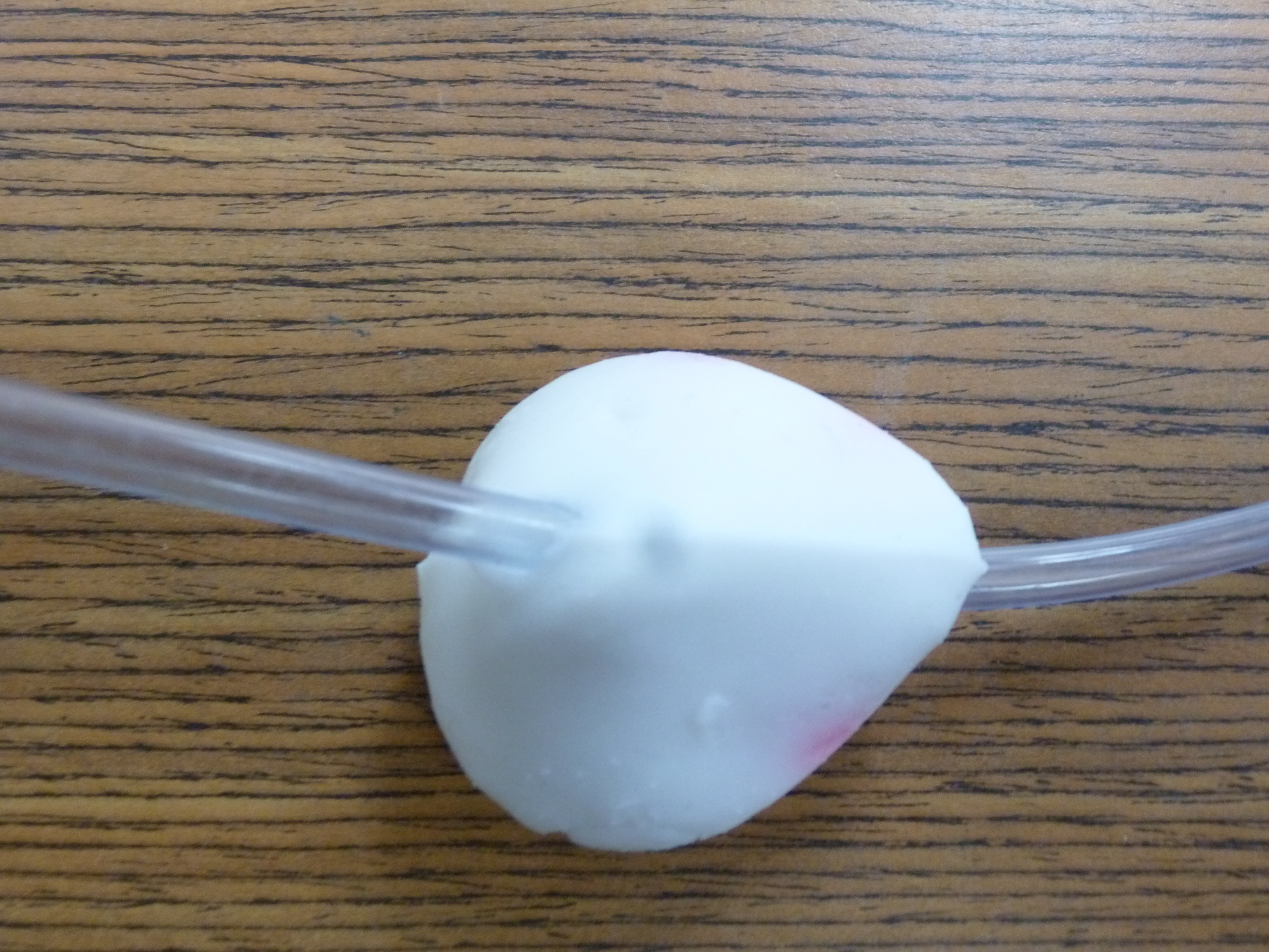
Prostate gland with visible lesion (pink)
with real a catheter tube replacing the urethra
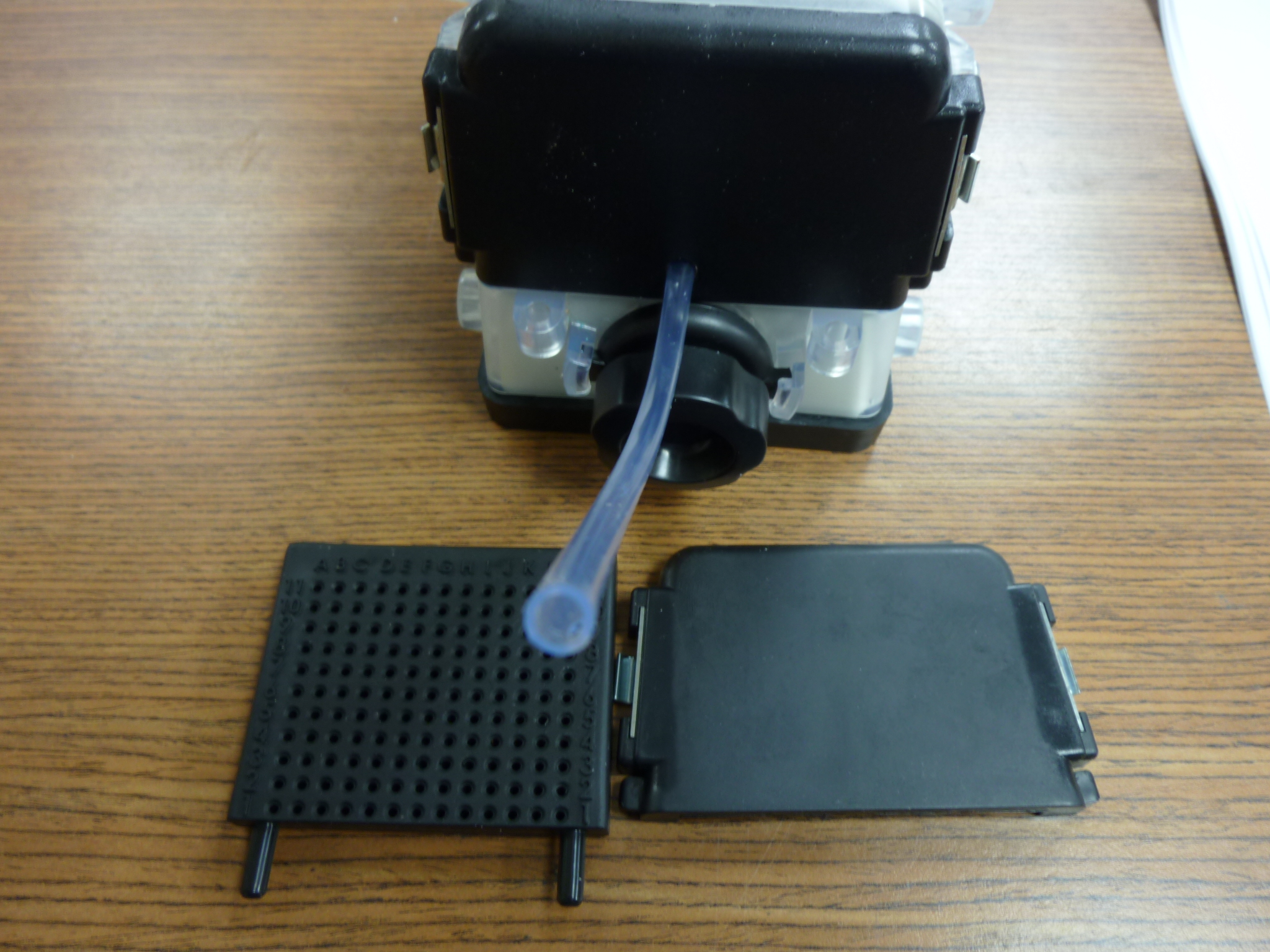
The Catheter Tubing coming out of the
removable front window through a matching hole
Catheter French Size: 18Fr.
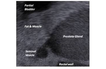
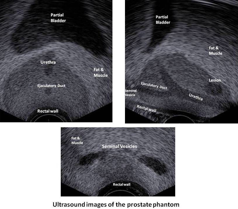
Ultrasound sagittal view of the prostate phantom
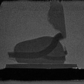
Sagittal MRI view of
the Prostate Phantom
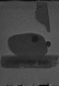
Sagittal MRI view of
the Prostate Phantom
Prostate Phantom
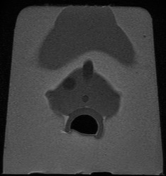
MRI Transverse
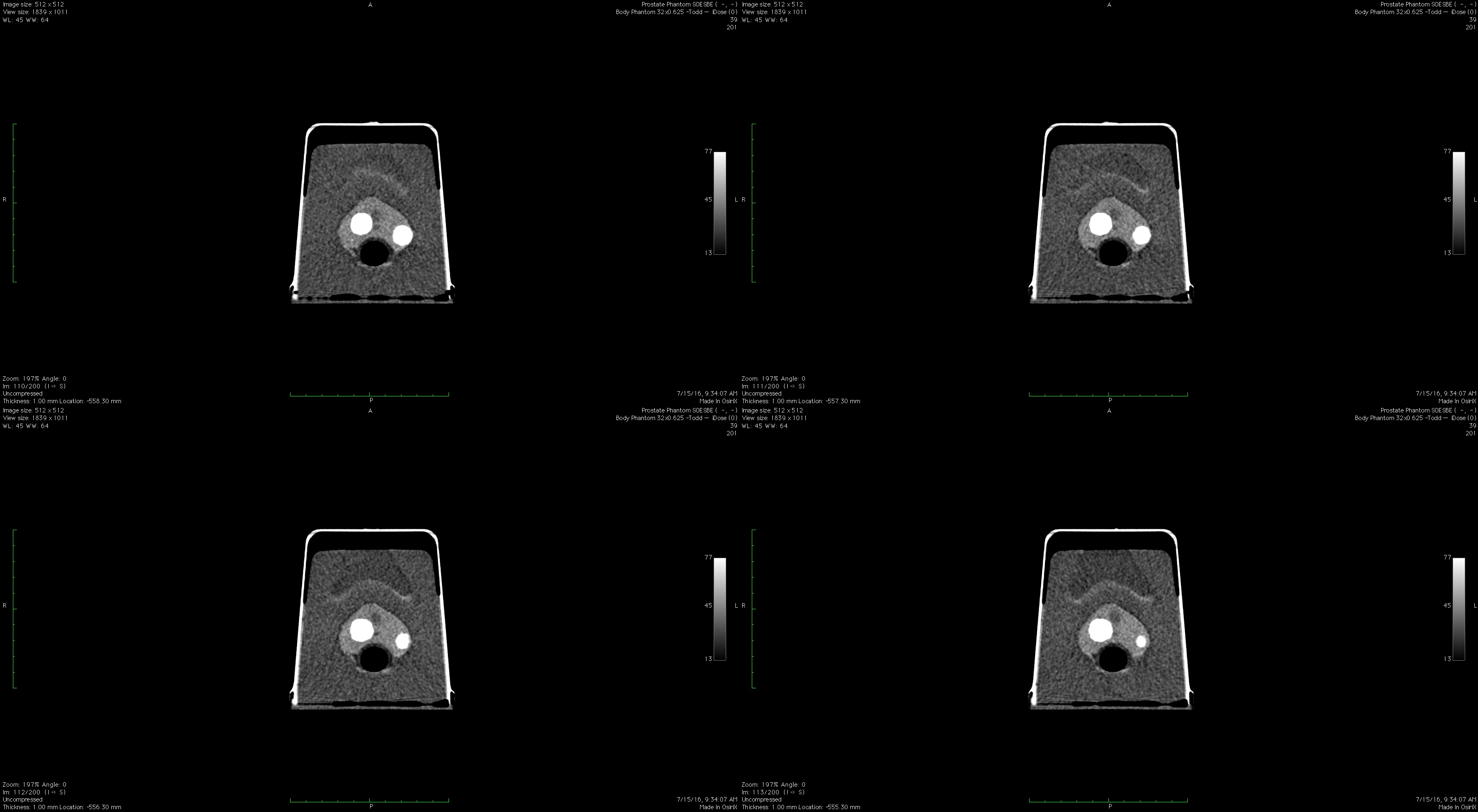
Prostate Phantom CT Transverse Scans
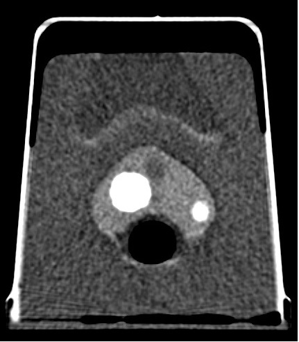
Enlarged Prostate Phantom CT
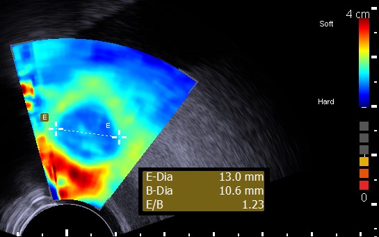
Prostate Phantom Elastography
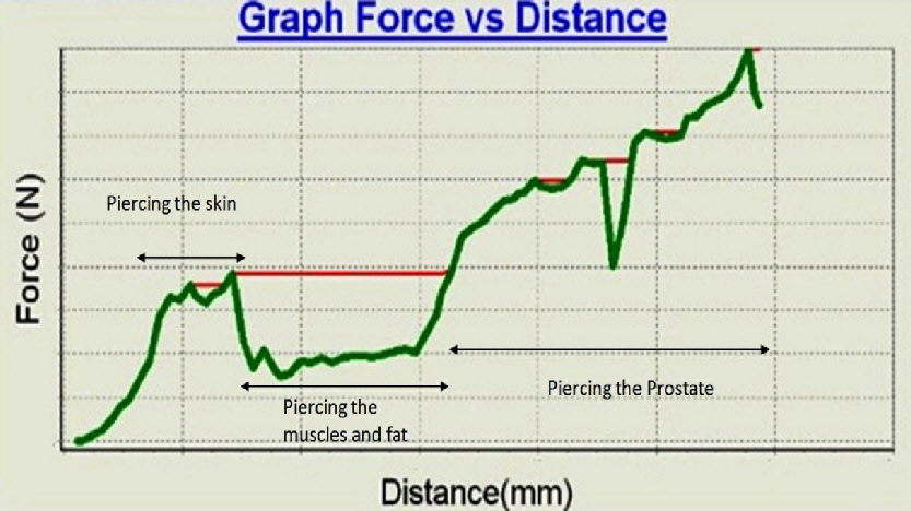
Real time graph (Prostate phantom device test bed) - of force as function of needle depth penetration.
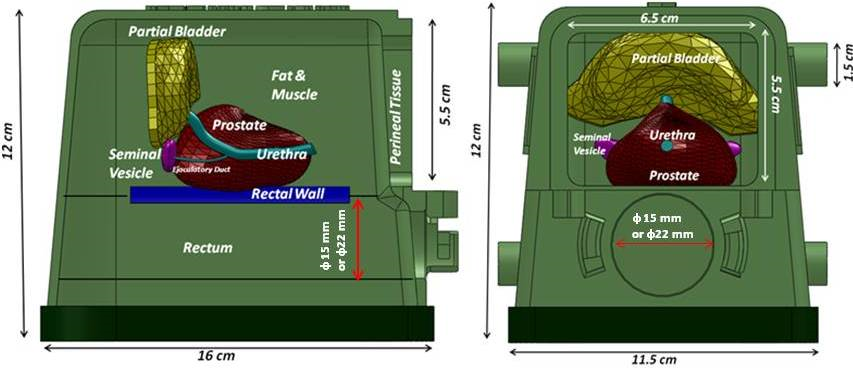
Internal tissues setup of the Brachytherapy prostate phantom. Diameter of 15 mm or 22 mm according to client demand.
Specifications :
Multi-layer material : each tissue or organ is independent and has its own characteristics defined by a real 3D shape, echogenicity level and mechanical properties.
Multiple usage of the same packaged phantom over an extended period of time.
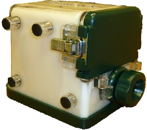
Enclosure :
16(L) x 11.5(W) x 12(H), Material . PVC, PC and metal latches. Front upper window 6.5(W) x 5.5 (H), Probe input diameter . 3.5 (all units in cm)
Perineal Tissue :
65(W) x 55 (H) x 3mm thick, approximate mechanical response of human tissue
Fat & Muscles :
Approximate mechanical response of human tissue
Urethra :
6mm diameter and 61mm(L)
Ejaculatory duct :
4mm diameter 28mm (L)
Seminal vesicles :
2 of 25(L)x6(W)x 4mm(Thick)
Prostate gland :
40cc, approximate mechanical response of human tissue.
Rectal wall :
81(L)x 75(W)x2.5(thick)mm, approximate mechanical response of human tissue.
Partial bladder :
13.4cc
Lesions :
4 Elliptic 0.3cc
Template:
13 columns and 11 rows.
ø: 2 mm